How the AIM Lab is transforming medical education
 In 2017, Dr. Matthew Bramlet, director of the AIM Lab and his team of engineers developed software capable of translating digital formats of medical scans into virtual reality for medical decision making, pre-surgical planning and patient education. The idea was to eliminate the need to 3D print a physical model and reduce the time it takes to view a complete image. It was also Dr. Bramlet’s belief that VR would enable clinicians to explore and experience the anatomy in ways they’ve never been able to before.
“We had some people that blew it up to the size of a cave and would walk through and orient themselves to the anatomy and then have that moment where everything sort of clicked into place in their mind,” said Dr. Bramlet who is also a pediatric cardiologist at the OSF HealthCare Children’s Hospital of Illinois. “Others would keep it down to the size they were looking for and cut through it with cut planes. Since it wasn’t a physical model, there were infinite opportunities for them to look into it.”
通过观察这些临床医生在VR中与3D解剖模型的互动,他注意到他们都在本能地讲述他们在虚拟空间中看到的东西,并分享沿途的发现。这时AIM实验室意识到他们创造了一种新的医学教育形式。随之而来的是endvo的商业衍生产品,这个平台允许临床教育者在虚拟现实中建立讲座,使用的仅仅是3D解剖模型、视频剪辑和图表。目前,政府、制造业和工业客户都在使用VR软件。
Tackling medical school training challenges with VR
 Before the first cases of COVID-19 hit the United States, the AIM Lab was working with researchers from the University of Illinois College of Medicine Peoria (UICOMP) and the Jump Trading Simulation & Education Center to teach medical students and health professionals outside of the hospital or classroom.
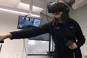
“Medical students can’t always participate in bedside rounds, assist in surgeries or learn from real-life scenarios where they have to diagnose critically-ill patients,” said Dr. Matthew Bramlet, director of the AIM Lab. “So, we wanted to investigate if we could give our students that same experience using virtual reality.”
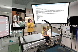
Dr. Bramlet and other researchers, including Dr. Teresa Riech built two cases in VR where medical students would have to diagnose and treat an adult with an unstable heart rhythm and a child who had difficulty breathing. The team then tasked 20 volunteer learners to go through each case at their own pace. Following the VR study, results showed:
- Learners rated VR equal to or better than traditional lecture.
- Medical students took the opportunity to review the material carefully, remaining in the learning environment well beyond recorded time (not an option in traditional lecture).
- The Immersive nature minimizes distraction. Not one student checked their phone to answer a text or look at a notification while wearing the VR headset.
- 由不同教师分别记录的案例,最终形成8秒内的模块,说明该模板可用于未来的案例开发。
The training also helped build students’ confidence. Before the lessons, only 5% expressed complete confidence in their ability to assess and provide the right treatment. 45% reported feeling completely confident in their skills after the VR lessons, 55% of students felt somewhat confident, and no students rated themselves as ‘not at all confident’. This research was presented at the Association of American Medical Colleges 2020 Annual Meeting.
Using VR to fight cancer
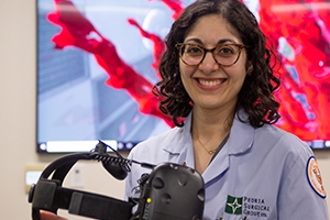 Surgical oncologist Dr. Sonia Orcutt first learned about the 3D modeling capabilities of Jump AIM through her husband, who is a pediatric cardiologist alongside Dr. Bramlet.
“就像儿科心脏外科医生一样,我们面临着同样的挑战,在我们到达手术室之前,我们不知道在手术室里会看到什么,”奥克特医生说。“所以,这激发了我的兴趣,我想研究使用VR来观察复杂的腹部肿瘤,以便进行术前规划的可行性。”
Dr. Orcutt worked with Dr. Bramlet to convert CT scans of previous surgical cases into VR and discovered that viewing the images in this technology would’ve helped her better define her operating plans. She now regularly uses VR to view complex cases where she can’t distinguish the relationship of a tumor with other structures, such as blood vessels or other major organs.
In one situation, she and her colleague were having a difficult time determining a plan of attack on a large tumor that was displacing a patient’s liver, inferior vena cava and kidney.
“When we placed our images in VR, we got a better understanding of the size and location of this tumor,” said Dr. Orcutt. “We ended up changing our incision to be a little larger, so we could have control of the blood vessels. We didn’t want to run into any significant hemorrhaging issues or anything else that was unsafe.”
Thanks to continued funding for the VR program, Dr. Orcutt is working on a formal study on whether the technology, in addition to standard imaging, improves a physician’s ability to define the extent of how much tissue should be cut out in a surgery. This can open the door to using this technology for a variety of tumors beyond the abdominal cavity.